 Rachel Sanders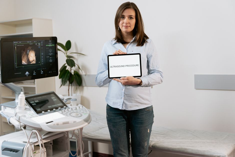
Your dog just had X-rays, and the vet comes back into the room saying, “I think we need to do an ultrasound.” Your stomach drops a little. You nod, but you’re already running through questions you don’t quite know how to ask. What are they looking for that the X-rays couldn’t show? Is this serious? How much is this going to cost?
You’re not alone in that moment. I’ve watched it happen hundreds of times in exam rooms. The word “ultrasound” carries weight, partly because most of us associate it with human pregnancy scans or with finding something wrong. Here’s what I tell people when they’re standing right where you are: an ultrasound is one of the most useful, least invasive diagnostic tools in veterinary medicine. Understanding what it actually does, and why your vet is recommending it, will make the whole experience far less stressful for both of you and your pet.
What a Pet Ultrasound Actually Does (and Why X-Rays Aren’t Enough)
X-rays are excellent at showing dense structures. Bones, foreign objects, the overall size and position of organs. But here’s the limitation: they produce a flat, two-dimensional image, and soft tissue detail gets lost in the process. Your vet can see that a kidney is enlarged on an X-ray, but they can’t tell whether there’s a cyst, a mass, a stone, or inflammation without looking inside it.
Ultrasound works differently. It uses high-frequency sound waves, usually in the range of 5 to 15 megahertz for small animals, that bounce off internal structures and return to a sensor. The machine converts those returning waves into a real-time image. You can actually watch the heart beating, see blood moving through vessels, and observe the texture of liver or spleen tissue in ways that simply aren’t possible with radiographs.
This is why ultrasound and X-rays are often used together. They answer different questions. A chest X-ray might show fluid around the heart. The ultrasound shows the heart itself, whether the chambers are functioning normally, and whether the fluid is coming from inside or outside the cardiac sac.
What Conditions Vets Use Ultrasound to Diagnose
The range is genuinely broad. In my experience, the most common reasons a vet will reach for an ultrasound include:
Abdominal concerns. This is probably the most frequent use. Vomiting, diarrhea, unexplained weight loss, blood in the urine, or abnormal bloodwork values will often prompt an abdominal ultrasound to look at the liver, spleen, kidneys, bladder, adrenal glands, and intestinal walls. A skilled sonographer can identify bladder stones as small as a few millimeters, detect splenic masses before they rupture, or spot the thickened intestinal walls associated with inflammatory bowel disease.
Cardiac evaluation. An echocardiogram is simply a cardiac ultrasound. It’s the gold standard for diagnosing conditions like dilated cardiomyopathy in Dobermans, mitral valve disease in Cavalier King Charles Spaniels, and hypertrophic cardiomyopathy in cats. No other tool gives you a real-time view of how the heart is actually pumping.
Pregnancy confirmation and monitoring. Unlike X-rays, which expose tissue to radiation, ultrasound is safe for pregnant animals and can confirm pregnancy as early as 21 to 25 days after breeding.
Guided procedures. This is the part that often surprises people. Ultrasound isn’t just diagnostic, it’s also used to guide needles precisely during fine-needle aspirates or biopsies. Your vet can watch the needle tip in real time and avoid major vessels, which makes the procedure significantly safer.
Endocrine disease. Conditions like Cushing’s disease cause the adrenal glands to enlarge. Ultrasound can visualize those glands and help distinguish between pituitary-dependent Cushing’s and an adrenal tumor, which changes the treatment plan entirely.
What to Expect on the Day of the Ultrasound
Knowing the logistics ahead of time genuinely reduces anxiety.
Fasting. For abdominal ultrasounds, your vet will typically ask you to withhold food for at least 8 to 12 hours beforehand. Food in the stomach and gas in the intestines scatter sound waves and degrade image quality significantly. Water is usually fine.
Fur clipping. A small area of fur will be shaved. Sound waves don’t travel well through air, and the fur traps air pockets between the skin and the probe. This surprises some owners. It grows back quickly.
Warm gel application. You’ve seen this on human scans. The gel eliminates air between the probe and the skin. It’s water-soluble and wipes off easily.
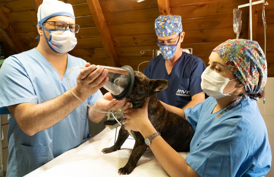
Positioning. Most pets are gently placed on a padded table, often on their back in a foam trough. Many dogs and cats tolerate this extremely well without sedation. Anxious animals, cats in particular, or cases where a biopsy is being performed may receive a mild sedative.
The scan itself. A typical abdominal ultrasound takes about 20 to 45 minutes depending on what’s being evaluated and how cooperative your pet is. You may or may not be allowed in the room, depending on your vet’s protocol.
Results. Your vet may review images with you immediately, or if the scan is being read by a board-certified radiologist (which provides greater diagnostic accuracy), results may come back within 24 to 48 hours.
General Practice Ultrasound vs. Specialist Referral: Which Do You Need?
| Feature | General Practice Ultrasound | Veterinary Internal Medicine Specialist |
|---|---|---|
| Who performs it | General practice vet, often self-trained | Board-certified internist or radiologist (DACVIM or DACVR) |
| Best for | Routine checks, bladder stones, pregnancy, obvious masses | Complex cardiac disease, oncology staging, guided biopsies, ambiguous findings |
| Availability | Most clinics | Specialty hospitals, university teaching hospitals |
| Image quality | Variable, depends on equipment and operator experience | High, with advanced equipment |
| Wait time | Usually same day or next day | 1-3 weeks typical |
| Feature | General Practice Ultrasound | Veterinary Internal Medicine Specialist |
|---|---|---|
| Who performs it | General practice vet, often self-trained | Board-certified internist or radiologist (DACVIM or DACVR) |
| Best for | Routine checks, bladder stones, pregnancy, obvious masses | Complex cardiac disease, oncology staging, guided biopsies, ambiguous findings |
| Availability | Most clinics | Specialty hospitals, university teaching hospitals |
| Image quality | Variable, depends on equipment and operator experience | High, with advanced equipment |
| Wait time | Usually same day or next day | May require referral, 1-7 days |
| When the AAHA standard matters | General practice hospitals meeting AAHA accreditation standards follow stricter equipment and protocol guidelines | Specialists follow their own board certification standards |
The honest answer is that many general practitioners do excellent ultrasound work, especially for straightforward cases. But if your vet finds something concerning or unusual, don’t hesitate to ask whether a specialist referral makes sense. A board-certified radiologist reviewing the images can catch things that a generalist might miss, and that second set of expert eyes can genuinely change outcomes.
How to Prepare and What to Ask Your Vet
A few questions worth writing down before the appointment:
- Will my pet need sedation, and what will that involve?
- Who will be reading the images, and how will I receive results?
- If something is found, what are the likely next steps?
- Is this an emergency ultrasound, or can it be scheduled within a few days?
The American Veterinary Medical Association recommends that pet owners actively participate in their pet’s care decisions, and asking these questions is exactly that kind of participation. You’re not being difficult. You’re being a good advocate.
If your pet has ongoing health concerns, having a basic pet first aid kit on hand at home is always smart (this site may earn a commission from qualifying purchases, and you should always confirm any home care steps with your vet first).
The goal of any diagnostic tool, ultrasound included, is simply to get you better information so you can make good decisions for your pet. It’s not the scary thing in the room. It’s actually one of the kinder ones: no needles, no radiation, just sound waves and a clear picture of what’s going on inside. You’ve already done the hard part by paying attention and getting your pet to the vet. The rest is just information, and information is almost always better than not knowing.
Sources
- AAHA accreditation standards
- American Veterinary Medical Association
- pet first aid kit
- PetSafe Easy Walk No-Pull Dog Harness
- Nylabone Power Chew Durable Dog Chew Toys
Disclosure: As an Amazon Associate, we earn a small commission from qualifying purchases at no extra cost to you. We only recommend products that genuinely support the topics covered in this article.
- EVERLIT 95-Piece Vet-Approved Pet First Aid Kit (~$32), Vet-approved 95-piece kit for dogs and cats, covers cuts, burns, sprains, and emergencies until you can reach a vet.
- Nutramax Cosequin DS Joint Supplement for Dogs (132ct) (~$36), The #1 veterinarian-recommended joint supplement brand, clinically studied for reducing joint pain in dogs.
Recommended Resources
Disclosure: As an Amazon Associate, we earn a small commission from qualifying purchases at no extra cost to you. We only recommend products that genuinely support the topics covered in this article.
- EVERLIT 95-Piece Vet-Approved Pet First Aid Kit (~$32), Vet-approved 95-piece kit for dogs and cats, covers cuts, burns, sprains, and emergencies until you can reach a vet.
- Nutramax Cosequin DS Joint Supplement for Dogs (132ct) (~$36), The #1 veterinarian-recommended joint supplement brand, clinically studied for reducing joint pain in dogs.